Research Highlights
Welcome to the NeuroNest Research Highlights page! Here, we dive into the fascinating world of neuroscience and psychology, bringing you the latest discoveries and insights. We are committed to providing fresh content every week so check back regularly for the latest insights, lab spotlights, and field notes from NeuroNest.
Student Work: We accept and showcase student-led projects, senior theses, and student research!
Field Notes: Stay up-to-date with concise, accessible summaries of new or interesting research papers and scientific breakthroughs in neuroscience and psychology. We deliver the key information you need, without the jargon.
Field Notes: Neurobiological Correlates of BPD
These researchers used graph analysis of resting-state fMRI data to reveal that individuals with Borderline Personality Disorder (BPD) exhibit significant disruptions in brain functional connectivity compared to healthy controls. Globally, BPD patients showed lower overall connectivity, while locally they demonstrated reduced centrality in specific limbic and temporal regions including the amygdala, hippocampus, and middle temporal gyrus, alongside increased centrality in the thalamus and anterior cingulate cortex. Importantly, the researchers linked these brain patterns to specific dimensional traits: the facet of separation insecurity correlated with global network efficiency, and depressivity in female patients was specifically associated with connectivity changes in the left middle temporal gyrus.
These findings suggest that the core psychopathological features of BPD are rooted in distinct neurobiological signatures, where specific network impairments may explain the intense fear of abandonment and social interpretation difficulties characteristic of this disorder. By identifying these correlations, the study emphasizes the potential for using functional connectivity measures as biomarkers for early identification, especially during late adolescence or early adulthood when interventions are most effective. Ultimately, this research supports a shift toward a dimensional approach to diagnosis (using tools like the PID-5), which could allow for more personalized and targeted psychiatric treatments based on an individual's unique neurobiological and personality profile.

Figure 1 Graphical representation of nodes with altered degree (top) and strength (bottom). The node size indicates greater statistical significance of the alteration. L left, R right, AMY amygdala, CAU caudate, IT inferior temporal, ParaC paracentral, THA thalamus, cAC caudal anterior cingulate, MT middle temporal, PERIC pericalcarine, HIP hippocampus, LING lingual, TP temporal pole, ENTH entorhinal, LOC lateral occipital, FP frontal pole, PC posterior cingulate.

Figure 2 Scheme of the functional connectivity network construction. The timeseries was extracted for each ROI, and Pearson’s correlation between the ROI timeseries was subsequently calculated to define the connection (matrix element) between the ROIs. By calculating this quantity for each pair of ROIs, a functional connectivity (FC) matrix for each patient was obtained. Finally, the FC matrices were proportionally thresholded in the range of 5–50% of the network density.
D’Adda, Francesca, et al. “Neurobiological Correlates of Personality Dimensions in Borderline Personality Disorder Using Graph Analysis of Functional Connectivity.” Scientific Reports, vol. 15, no. 1, 12 Apr. 2025, www.nature.com/articles/s41598-025-85989-x, https://doi.org/10.1038/s41598-025-85989-x. Accessed 9 June 2025.
Field Notes: Taste and Color

Figure 3 Violin plot visualizing the distribution and density of the data. The horizontal lines within the violin plot represent the median of the data. Error bars indicate the standard deviations. The shapes of the violins capture the density of the data at different values, with wider sections indicating a higher density of data points; the faint grey lines indicate the range of the data, showing the minimum and maximum values. Mean proportion of binding errors (BEs) for each taste pair (sweet/sour, sweet/salty, sour/salty) under congruent and incongruent conditions. The graphs clearly show that binding errors are significantly more frequent in the incongruent conditions than in the congruent conditions for all taste pairs, suggesting a robust effect of color-taste congruency on visual feature binding. The figure shows that participants made significantly more ICs in the incongruent condition than in the congruent condition for each taste pair. For the Sour/Salty pair, the rates were 7.35 % vs. 2.92 %; For the Sweet/Salty pair, the rates were 6.84 % vs. 3.26 % and For the Sweet/Sour pair, the rates were 6.34 % vs. 3.00 %.
Bortolotti, Alessandro, et al. “Color–Taste Correspondences Influence Visual Binding Errors.” Acta Psychologica, vol. 254, Apr. 2025, p. 104785, https://doi.org/10.1016/j.actpsy.2025.104785. Accessed 9 June 2025.
This study investigated how established color-taste correspondences, like associating sweetness with red, sourness with yellow, and saltiness with blue, influence the accuracy of visual feature binding. Using an experiment involving divided spatial attention, researchers found that participants were significantly more likely to make illusory conjunctions (binding errors where features are mismatched) when food words were presented in colors that were incongruent with their associated taste (like the word "sweet" in yellow font) compared to congruent pairings. Specifically, the rate of binding errors rose to 20.5% in incongruent trials compared to only 9.18% in congruent ones, whereas correct identifications (hits) were significantly higher when the color and taste associations matched. These findings demonstrate that visual feature binding is not a purely automatic, bottom-up process but is heavily modulated by top-down mechanisms, including prior knowledge and learned expectations. The paper suggests that these crossmodal associations may act as perceptual "priors" that shape the brain's predictions during early stages of visual processing or within short-term memory. Ultimately, this research supports an interactive model of perception, showing that information typically associated with one sense (taste) can fundamentally bias how we perceive and integrate the features of another (vision).
Field Notes: Linguistics & Early Psychosis Detection
The Accelerating Medicines Partnership in Schizophrenia (AMP SCZ) initiative demonstrated that automated analysis of language, acoustics, and facial expressions can serve as a scalable, objective biomarker for early psychosis detection. By analyzing data from individuals at clinical high risk (CHR) compared to community controls, these researchers found that CHR individuals consistently displayed a significant overuse of pronouns, extending across personal, possessive, and third-person forms, alongside a decrease in descriptive content words such as adjectives, adverbs, and nouns. While CHR participants appeared to show higher syntactic complexity in some settings, the study found this was typically a byproduct of increased verbal output in structured tasks rather than an inherent grammatical trait. These findings imply that emerging psychosis involves a linguistic shift away from words with inherent meaning toward context-dependent language, reflecting deeper disruptions in perspective-taking and discourse structure.

Figure 6 ORs for grammatical features, syntactic dependencies, and parts of speech across three language sample types—PSYCHS interviews (blue), open-ended interviews (green), and audio diaries (audio). An OR of 1 indicates no association with CHR status. ORs greater than 1 suggest that higher feature values are associated with increased odds of being classified as CHR, while ORs less than 1 indicate a negative association. Features marked with an “x” were statistically significant based on the Wald z-test.
Bilgrami, Zarina R, et al. “Collecting Language, Speech Acoustics, and Facial Expression to Predict Psychosis and Other Clinical Outcomes: Strategies from the AMP® SCZ Initiative.” Schizophrenia, vol. 11, no. 1, 15 Oct. 2025, pp. 125–125, https://doi.org/10.1038/s41537-025-00669-z. Accessed 7 Jan. 2026.
Field Notes: Vegetative States and Consciousness

Figure 3 Results of two sample communication scans obtained from Patient 23 (Panels A and C) and a healthy control subject (Panels B and D) during functional MRI are shown. In Panels A and B, the observed activity pattern (orange) was very similar to that observed in the motor-imagery localizer scan (i.e., activity in the supplementary motor area alone), indicating a “yes” response. In Panels C and D, the observed activity pattern (blue) was very similar to that observed in the spatial-imagery localizer scan (i.e., activity in both the parahippocampal gyrus and the supplementary motor area), indicating a “no” response. In Panels A and C, the name used in the questions have been changed to protect the privacy of the patient.
Monti, Martin M., et al. “Willful Modulation of Brain Activity in Disorders of Consciousness.” New England Journal of Medicine, vol. 362, no. 7, 18 Feb. 2010, pp. 579–589, https://doi.org/10.1056/nejmoa0905370.
This study focused on the willful modulation of brain activity in disorders of consciousness using fMRI methods. The researchers sought to assess residual awareness in 54 patients diagnosed with vegetative or minimally conscious states (MCS), given the high rate of clinical misdiagnosis (approximately 40%) in this population. The main finding was that 5 of the 54 patients were able to willfully modulate their brain activity by generating neuroanatomically specific responses when instructed to perform mental imagery tasks: motor imagery (imagining playing tennis, activating the supplementary motor area) and spatial imagery (imagining navigating a city, activating the parahippocampal gyrus). Four of these five patients had been admitted with a diagnosis of being in a vegetative state, demonstrating that they retained awareness and cognitive function despite meeting the behavioral criteria for unresponsiveness. This study's biggest implication is that fMRI can reveal covert awareness when traditional bedside assessments, which rely on motor responses, fail. The researchers also developed a technique to use this willful brain modulation for functional communication. One patient (Patient 23), who remained behaviorally unresponsive despite thorough clinical reassessment, was able to answer yes/no questions accurately by selectively activating the supplementary motor area for one answer (yes) or the parahippocampal gyrus for the other (no). For five of the six questions posed, the patient provided the factually correct answer. These results ultimately show that a small proportion of unresponsive patients have intact awareness and cognition, and this fMRI technique may be useful for detecting this consciousness and establishing basic interactive communication with patients who cannot move or speak.
Field Notes: The Neural Correlates of Dreaming - Challenging Prior Work
This study identified a parieto-occipital “hot zone” as the core neural correlate of conscious experience, or dreaming, during sleep. Researchers used high-density EEG during serial awakenings in both Non-REM (NREM) and REM sleep to contrast dream experiences (DE) with no experiences (NE). They found that dreaming was consistently associated with a local decrease in low-frequency EEG activity (1-4 Hz) and a simultaneous increase in high-frequency activity (20-50 Hz) in this posterior region, which includes the medial and lateral occipital lobe, the precuneus, and the posterior cingulate gyrus. The presence of specific dream contents, such as faces, speech, or thought-like experiences, correlated with increased high-frequency activity in corresponding specialized cortical areas, mirroring brain activity seen during wakeful perception. This localized bispectral activity allowed the researchers to predict, with 87% total accuracy, whether a subject would report dreaming during NREM sleep. These findings resolve the paradox that dreaming can occur in both NREM sleep and REM sleep, by demonstrating that consciousness requires only the localized activation of this posterior hot zone, regardless of the overall global EEG state. This localization challenges the traditional notion that the neural correlate of consciousness corresponds to a broad frontoparietal network, suggesting the core correlate during sleep could be restricted to posterior regions. The ability to predict consciousness in real time based on this localized activation suggests that this technique could be useful in future work on disorders of consciousness to detect awareness when patients lack behavioral responsiveness.

Figure 1 Dreaming experience vs. no experience in NREM sleep (low- frequency power). (a) Cortical distribution of t values for the contrast between DEs and NEs at the source level for low-frequency power (1–4 Hz) in NREM sleep (20 s before the awakening). P < 0.05, after correction for multiple comparisons (two-tailed paired t-tests, n = 32 subjects, t31 > 2.04. (b) Same as a but for the contrast between NE and DEWR (two-tailed paired t-tests, n = 20 subjects, t19 > 2.09). LL, left lateral view; RL, right lateral view; LM, left medial view; RM, right medial view.

Figure 4 The content of dream experiences in REM sleep. (a) Correlation between the thinking–perceiving score and 25–50-Hz power (last 8 s, n = 7 subjects). Left: mean Spearman rank correlation coefficients (n = 7 subjects). Right: significant voxels (P < 0.05, one-tailed permutation test, r > 0.14). (b) Left: 25–50-Hz power differences (DE with face minus DE without face). ROI contrast in the fusiform face area (FFA) (P = 0.023; one-tailed paired t-test, n = 7 subjects, t6 = 2.52). Right: FFA (red). (c) Upper row: 25–50 Hz average power differences between DEs with and without a spatial setting (n = 6 subjects, t5 > 2.57). Right: right posterior parietal cortex. Middle row: movement vs. no movement (n = 7 subjects, t6 > 2.45). Right: superior temporal sulcus. Bottom row: speech vs. no speech (n = 7 subjects, t6 > 2.45). Right: Wernicke’s area. Two-tailed paired t-tests; P < 0.05 (red) and P < 0.1 (yellow).
Siclari, Francesca, et al. “The Neural Correlates of Dreaming.” Nature Neuroscience, vol. 20, no. 6, 10 Apr. 2017, pp. 872–878, www.nature.com/articles/nn.4545, https://doi.org/10.1038/nn.4545.
Field Notes: The Gorilla Strikes Again!
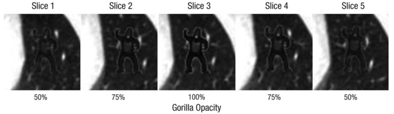
Figure 1: Illustration of the slices showing the gorilla in the final trial of Experiments 1 and 2. The opacity of the gorilla increased from 50% to 100% and then decreased back down to 50% over the course of 5 slices within a stack of 239.
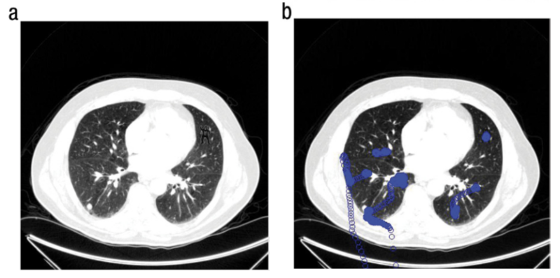
Figure 2: Computed-tomography image containing the embedded gorilla (a) and eye-position plot of a radiologist who did not report seeing the gorilla (b). In (b), the circles represent eye positions recorded at 1-ms intervals.
**DO YOU SEE THE GORILLA? Look to the top right corner of Panel A.
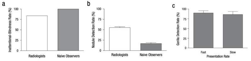
Figure 3: Experimental results. The graph in (a) shows the rate of inattentional blindness (i.e., the percentage of observers who did not report seeing the gorilla) among the radiologists in Experiment 1 and the naive observers in Experiment 2. The graph in (b) shows the percentage of nodules that were correctly marked by these same observers. The graph in (c) shows the rate at which observers in Experiment 3 detected the gorilla as a function of presentation rate (fast: 35 ms/frame; slow: 70 ms/frame). Error bars represent standard errors of the mean.
This paper investigates the phenomenon of sustained inattentional blindness, where people often miss a salient, unexpected event when they are deeply engaged in a different task, an effect popularized by the "Invisible Gorilla" demonstration (see attached video below). The key background question addressed was whether highly specialized experts, such as radiologists who spend years honing their visual detection skills, are still vulnerable to inattentional blindness while performing complex searches within their own domain. To test this, 24 expert radiologists performed their familiar task of searching through hundreds of CT slices of the lung for small nodules. In the final trial, the researchers inserted a large, highly anomalous stimulus- a gorilla outlined in white, measuring more than 48 times the size of an average nodule. The primary results were striking: 83% (20 of 24) of the expert radiologists failed to report seeing the gorilla. Furthermore, eye-tracking data revealed that the majority of those who missed the gorilla looked directly at its location. Although the experts were significantly better at the primary nodule detection task than naive observers (who missed the gorilla 100% of the time), their high miss rate demonstrates that even expert searchers are not immune to inattentional blindness. The implications of these findings are that high levels of expertise do not overcome the inherent limitations of human attention and perception. Since observers were only focused on searching for small nodules and were not expecting an item like a gorilla, their focused attention acted like "blinders".
Drew, T., Võ, M. L.-H., & Wolfe, J. M. (2013). The invisible gorilla strikes again. Psychological Science, 24(9), 1848–1853. https://doi.org/10.1177/0956797613479386
Click below to try out the original gorilla experiment and experience inattention blindness first hand!
Field Notes: Facial Perception & Differentiation of In vs. Out Groups
Researchers investigated the early perceptual underpinnings of the racial outgroup homogeneity effect, where individuals tend to individuate members of their own group (ingroup) but process members of other groups (outgroups) categorically. The research aimed to determine if this deindividuation is a mistake in later recollection or if it emerges in the initial stages of sensory perception. Using an fMRI adaptation paradigm, self-identified White participants viewed blocks of White and Black faces that parametrically varied in physical similarity. The core finding revealed that racial disparities emerge in the tuning of the face-selective cortex. Specifically, other-race (Black) faces elicited a more gradual and smaller release from adaptation compared with own-race (White) faces, particularly in the right face-selective cortex. This neural pattern suggests that the face-selective cortex is narrowly tuned toward the individual identity of ingroup members but is more broadly tuned toward social category information for outgroup members, leading to habituation to dissimilar outgroup targets as if they were repeated instances of the same identity. They also found that the volume of face-selectivity in the bilateral ventral temporal cortex was significantly lower when responding to Black faces. These neuroimaging results paralleled behavioral findings showing that participants individuated own-race faces to a greater extent across measures of perceived similarity, discrimination, and recognition memory. The conclusion is that biases for other-race faces emerge at some of the earliest stages of sensory perception, providing a fine-grained mechanism that may precede subjective experience and potentially cascade into harmful outcomes like the application of stereotypes or faulty eyewitness testimony.
Hughes, B. L., Camp, N. P., Gomez, J., Natu, V. S., Grill-Spector, K., & Eberhardt, J. L. (2019). Neural adaptation to faces reveals racial outgroup homogeneity effects in early perception. Proceedings of the National Academy of Sciences, 116(29), 14532–14537. https://doi.org/10.1073/pnas.1822084116

Figure 1: The volume of face-selectivity in high-level visual cortex is modulated by face race. (A) Thresholded parameter maps show voxelwise t values for the contrast of White faces (Top) or Black faces (Bottom) versus all other stimuli. Data are presented on inflated cortical surfaces of 3 participants (dark regions are sulci; lighter regions are gyri). The region shown is the ventral surface of the temporal lobe, known as the VTC. The blue region outlined in dotted-white is the MFS, whose posterior and anterior tips are anatomical anchors predicting the location of face-selective cortex on the lateral fusiform gyrus. (B) Line plots illustrating the volume of above-threshold (t values > 3) activation in each subject’s VTC defined with the contrasts of White or Black faces.
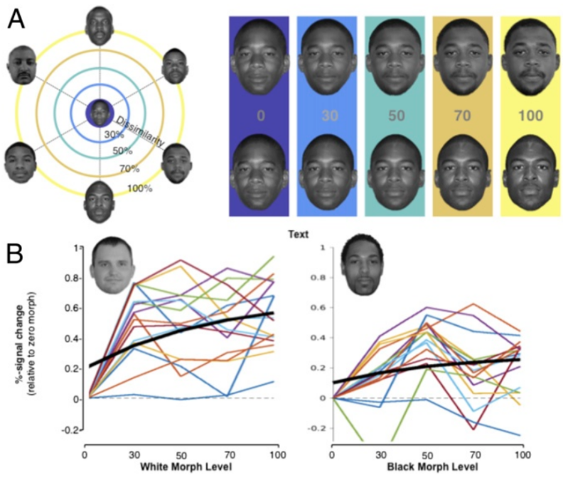
Figure 2: (A) Experimental design of the adaptation experiment. In each block, 6 faces were presented at 1 of 5 levels of groupwise dissimilarity. An example of the morphing scheme is presented on the Left, and 2 example morph lines are shown on the Right. (B) Plots mapping the percentage of signal change in right hemisphere face-selective cortex in each participant for White (Left) and Black (Right) faces, normalized to the zero-morph level (full adaptation), along with the summary curve in black.
Student Work: Neural Mechanisms of Choreographic Memory: An EEG Study on Dance Learning and Recall
While dance is fundamental to human culture, its neural basis is underexplored. This study examined dance learning and recall using EEG. Fifteen dancers watched seven short clips while wearing an EEG cap, followed by 30-second mental imagery periods. After all the clips, they performed the choreography from memory. Alpha power increased significantly during imagery, especially in posterior regions, suggesting its role in memory recall and motor planning. A strong positive correlation showed that individuals with higher alpha power during video watching also had higher alpha power during imagery. These findings highlight EEG’s potential to reveal choreographic memory processes. Future research should explore marking, verbal rehearsal, and other memory-enhancing techniques in dance learning.

Figure 2: Topological maps of mean alpha power during a) video watching and b) mental imagery conditions. c) The plot highlights electrodes with significant differences (p < 0.05) between conditions (PO4 and P8).

Figure 4: Comparison of Alpha Power During Video Watching and Mental Imagery Conditions. a) Raincloud plot. Individual participants’ alpha power values are plotted, with grey lines connecting paired values. Most lines trend upward, indicating higher alpha power during Mental Imagery. b) Boxplots. The median, interquartile range, and whiskers are displayed for each condition. Higher and more variable alpha power is evident during Mental Imagery. c) Density Plots. Overlapping distributions show a rightward shift for Mental Imagery, reflecting increased alpha power compared to Video Watching.
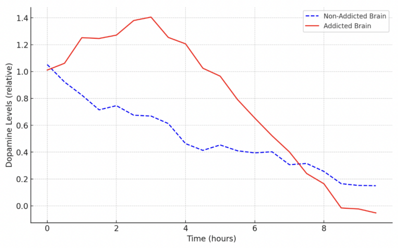
Figure 2. Dopamine Pathway Alterations in Addiction .
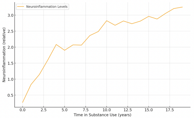
Figure 4. Neuroimmune Interaction In Addiction
Field Notes: New Frontiers in Addiction Treatment
Addiction is a complex brain disorder that changes how the brain’s reward, motivation, and control systems work. It’s not just about willpower- addiction rewires the brain at a molecular level, making it hard to quit. Scientists are now exploring new treatments that target these brain changes directly. Gene therapies like CRISPR-Cas9 aim to edit genes linked to addiction, while drugs called HDAC and DNMT inhibitors may help reactivate genes that addiction has turned off. Another important area is neuroimmune modulation: chronic drug use causes brain inflammation, which worsens addiction. Anti-inflammatory drugs and vaccines that block drugs from reaching the brain show promise in reducing cravings. Despite these advances, treating addiction remains difficult because it involves many factors, including genetics and environment. Emerging technologies like brain stimulation and digital health tools may help improve recovery in the future. Overall, these new approaches offer hope for more personalized and effective addiction treatments beyond just relying on willpower.

Table 3. Gene Therapy and Epigenetic Modulation Approaches in Addiction Treatment .
Aejeeliyah Yousuf. (2025). Neuroscience of Addiction Molecular Mechanisms and Potential Therapeutic Targets. Middle East Journal of Pure and Applied Sciences (MEJPAS), 1(1), 55-63. https://mideastjournals.com/index.php/mejpas/article/view/12
Field Notes: Trauma, Addiction, and the Quiet Power of Mindfulness
This study investigated how Adverse Childhood Experiences (ACEs) affect individuals with Opioid Use Disorder (OUD), particularly in relation to pain interference (how much pain disrupts daily life). People with high ACEs tend to experience more intense psychological symptoms and often respond poorly to standard treatments. The researchers examined whether mindfulness-based interventions (MBIs), which promote self-awareness and emotional regulation, could help this population by targeting two psychological processes linked to trauma and chronic pain: self-critical rumination (SCR), or the tendency to dwell on self-directed negative thoughts, and pain catastrophizing, which involves exaggerating the severity or impact of pain. They hypothesized that ACEs lead to increased SCR, which in turn increases pain catastrophizing and worsens pain interference. Using data from a 24-week clinical trial comparing a trauma-informed online mindfulness program (M-ROCC) to an active recovery support group, they found that ACEs influenced treatment outcomes only in the MBI group. In that group, reductions in SCR mediated reductions in pain catastrophizing, supporting the idea that MBIs can interrupt harmful psychological cycles in those with high ACEs. No significant group-level differences emerged, but the findings suggest that MBIs may offer particular benefits for individuals with trauma histories by addressing specific thought patterns that worsen pain and psychological distress.

Figure 1: Different temporal paths of symptom changes in the (A) M-ROCC and (B) active control group

Figure 2: Correlations between ACE scores and ΔSCRS at week 8 in the M-ROCC treatment arm.
Joss, D., Rosansky, J., Gardiner, P., Edwards, R. R., Weiss, R. D., Napadow, V., & Schuman-Olivier, Z. (2025). Modulating mechanisms of adverse childhood experiences in a mindfulness-based intervention: Preliminary insights from an opioid use disorder study. Frontiers in Psychology, 16. https://doi.org/10.3389/fpsyg.2025.1529106
Student Work: Hyperventilation and Blood Alkalization Effect on Seizures in a Mouse Model of Absence Epilepsy
Ashley Mchugh
B.S. in Cognitive Science, University of Virginia
Absence epilepsy is a childhood neurological disorder marked by brief lapses in consciousness, often seen as staring spells. These seizures can be triggered by hyperventilation, but the underlying mechanisms are not well understood. One theory suggests blood alkalization from hyperventilation may be the cause. This study tested whether hyperventilation induces spike-wave discharges (SWDs), the brain activity signature of absence seizures, in C3H/HeJ mice, a model of the disorder. EEG headsets were used to monitor SWDs during experiments using two setups: (1) a head-fixed rig with hypoxic gas delivery via nose cone, and (2) plethysmography chambers that better induced hyperventilation by adjusting gas composition. An additional experiment used acetazolamide, a drug that lowers blood pH, to test whether reversing alkalization affects SWDs. The head-fixed setup failed to reliably trigger hyperventilation, showing no change in SWDs. In contrast, the plethysmography chambers effectively induced hyperventilation, leading to increased breathing rate and SWD activity. The acetazolamide results were inconclusive due to small sample size and high variability.
Field Notes: ipER, Borderline Personality Disorder and Memory
Krause-Utz, A., Saygin, M., Podbylska, M., Chatzaki, E., la Rosa, B., & Lis, S. (2025). Interpersonal emotion regulation, borderline personality disorder symptoms, and working memory during social-affective distraction. Personality Disorders: Theory, Research, and Treatment, 16(3), 210–222. https://doi.org/10.1037/per0000722
This paper explores how emotion regulation (ER), particularly interpersonal emotion regulation (ipER), is influenced by context, especially in social situations where emotions signal mental states and secure support. While most research has focused on internal ER strategies, recent studies highlight the importance of ipER, where emotions are regulated through social interactions. Hofmann et al. (2016) identified four key ipER strategies: enhancing positive affect, perspective-taking, social modeling, and soothing.
In Borderline Personality Disorder (BPD), which involves heightened emotional sensitivity and difficulty returning to baseline emotions, individuals struggle with ipER, particularly under relational stress. People with BPD often use interpersonal support ineffectively, engage in maladaptive behaviors, and report lower emotion regulation efficacy. Cognitive deficits, especially in attention and working memory (WM), may hinder ipER by affecting responses to social-emotional cues.
The study found that poorer WM performance was linked to reduced use of ipER strategies, particularly enhancing positive affect. Higher BPD symptom severity also predicted less frequent use of ipER strategies. Working memory deficits partially explained these difficulties, suggesting emotional inflexibility, rather than the complete absence of strategies, as a core issue in BPD.
Field Notes: The Heart's Own "Little Brain"
This 2024 update highlights how neuroscience is becoming a game-changer in treating heart disease. It explores the role of the autonomic nervous system (ANS), the part of the nervous system that automatically controls things like heart rate and blood pressure, and how its dysfunction can contribute to serious cardiac conditions, including arrhythmias and sudden cardiac death.
What’s especially exciting is the paper’s focus on new therapies that use neuroscience to actually modulate heart function. For example, vagus nerve stimulation, spinal cord stimulation, and other neuromodulation strategies are being developed to "reset" how the brain and heart communicate. These treatments aim to calm overactive sympathetic responses (the "fight or flight" system) or strengthen weakened parasympathetic signals (the "rest and digest" system), offering more precise, personalized treatment options.
In short: the future of heart health might just be through the nervous system!

Figure 1: Organization of cardiac neural control
Ajijola, O. A., Aksu, T., Arora, R., Biaggioni, I., Chen, P., De Ferrari, G., Dusi, V., Fudim, M., Goldberger, J. J., Green, A. L., Herring, N., Khalsa, S. S., Kumar, R., Lakatta, E., Mehra, R., Meyer, C., Po, S., Stavrakis, S., Somers, V. K., … Shivkumar, K. (2025). Clinical neurocardiology: Defining the value of neuroscience‐based Cardiovascular therapeutics – 2024 update. The Journal of Physiology, 603(7), 1781–1839. https://doi.org/10.1113/jp284741
Field Notes: Can Parenting Shape Personality Into Adulthood?
A new longitudinal twin study suggests that affectionate parenting during childhood (ages 5-10) may positively shape personality traits like openness, conscientiousness, and agreeableness by the time teens reach adulthood at age 18. Using over 2,000 identical twins in the UK, researchers found that even small differences in parenting between twins led to measurable differences in these traits despite shared genetics and environment. While effects were modest, the findings support the idea that positive parenting has lasting, meaningful impacts on who we become, with potential for public policy and early intervention.
Wertz J, Moffitt TE, Blangis F, Ambler A, Arseneault L, Danese A, Fisher HL, Caspi A. Parenting in childhood predicts personality in early adulthood: A longitudinal twin-differences study. Am Psychol. 2025 Apr 17. doi: 10.1037/amp0001508. Epub ahead of print. PMID: 40244963.
Field Notes: GABA and Psychosis - It’s Not Just About One Brain Region
Disruptions in the GABA system- a key inhibitory neurotransmitter- may play a role in schizophrenia and early psychosis. While past studies have found reduced GABAergic neurons and receptor levels in the hippocampus, this new PET imaging study reveals something deeper: it's not just about changes in one brain region. Instead, individuals with first-episode psychosis or at high risk for psychosis show altered network-level organization of a specific GABA receptor (GABA A α5), even though the overall amount of this receptor in the hippocampus isn't significantly different from healthy controls. This suggests that psychosis may involve system-wide dysregulation in how GABA A α5 receptors are coordinated across the brain, not just local deficits.
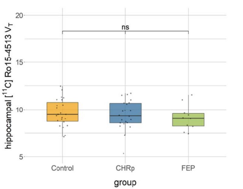
Figure 1: [11C]Ro15-4513 binding in the hippocampus between groups.

Figure 2: Pertubation covariance z-score statistics.
Lukow, P. B., Schubert, J. J., Severino, M., Knight, S. R., Kiemes, A., Livingston, N. R., Davies, J., de Micheli, A., Spencer, T. J., Fusar-Poli, P., Haege, B., Vorontsova, N., Donocik, J., Rabiner, E. A., Grace, A. A., Williams, S. C., McGuire, P., Veronese, M., Turkheimer, F. E., & Modinos, G. (2025). GABAaReceptor Availability in Clinical High-Risk and First-Episode Psychosis: A [11C]RO15-4513 Positron Emission Tomography Study. https://doi.org/10.1101/2025.02.27.25322861
Create Your Own Website With Webador